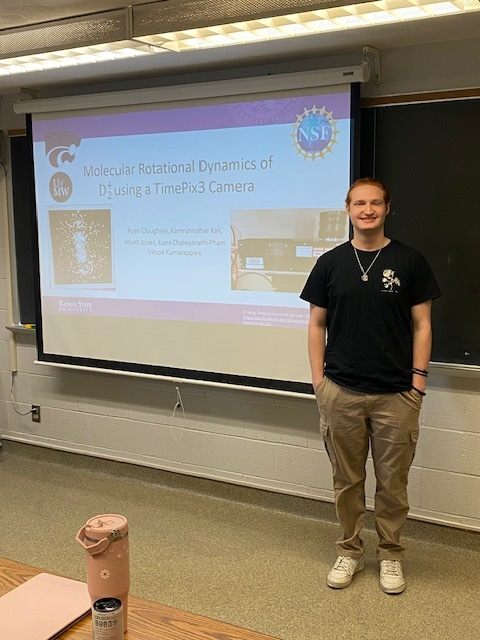
Molecular Rotational Dynamics of D2+ using a TimePix3 Camera
Ryan Cloughley, University of Mary Washington, Physics Major
Mentored by Dr. Vinod Kumarappan

The focus of our project is studying the rotational dynamics of D2+ using a TimePix3 (TPX3) camera. To do this, we use a pump-probe setup, seen in Figure 1, to excite D2 into D2+, then, after a short time delay, we dissociate the molecule. We then collect the fragments from the dissociated molecule, D+ and D, using a velocity map imaging (VMI) setup. The VMI setup uses microchannel plating to amplify the signal from ion species which then release an avalanche of electrons. These electrons excite a phosphor screen to emit photons, which the TPX3 camera detects.
The TPX3 is a 256x256 pixel camera that records time over threshold (TOT), time of arrival (TOA), and position information. Each pixel is 55 μm × 55 μm and measures TOT and TOA information on an event-driven basis [3]. Event-driven measurements mean that each pixel on the camera measures information independent of each other, without being limited by a shutter speed. Instead, the refresh rate of the pixels and the speed at which the phosphor screen dims limit our time resolution. Even with these limitations, the TPX3camera has a time resolution of 1.56 ns, which is enough to differentiate the time of flight of different ions [4]. This allows us to measure TOT, TOA, and position information about many ion species from a parent molecule without needing to gate the detector to isolate species [2].

Fig. 1. A diagram of the experimental setup
Our experimental setup, as seen in Figure 1, is a pump-probe experiment which uses ~40fs laser pulses at 800 nm for pumping and probing the molecule. The position of the stage determines the length of the probe arm, which allows us to control the time delay between exciting and probing the molecule. In our experiment, it is helpful to refer to the arms as the strong (upper arm) and weak (lower arm), since we measure on both sides of the temporal overlap of the lasers. When the weak pulse comes first, we align D2 , then dissociate the molecule with the strong pulse. When the strong pulse comes first, we ionize D2 creating D2+ ions, which we then dissociate with the weak pulse. Probing with the weak pulse is reasonable since ground electronic state of D2+ can be dissociated from by 1-3 photons [1]. Another important feature of our experimental setup is how we eliminate background signals. We do this by using the chopper and pulsed gas jet to control the measurements in the chamber. We measure four different statuses: pump-probe-gas, pump-probe, probe-gas, and probe. Using these statuses, we can normalize our signal to help eliminate background noise.
After processing the data that comes directly from the TPX3 camera, we have information about time of flight, position, and counts for each delay and status. Then, combining all this information, we can make plots to help us interpret our measurements. We use the time-of-flight graphs, as seen in Figure 2, to help identify ions so we can gate the data to look more closely at the ion species of interest. Then, we create delay vs ion yield, Figures 3, which allows us to investigate the rotational motion of our molecule. Finally, we make x vs y plots, Figure 4, which let us see symmetries and different kinetic energy levels present in our measurements.
 Fig. 2. Plots of the time of flight information collected by the pixels (top) and time of flight information collected by the pixels in each column (bottom).
 Fig. 3. Plots for the alignment of D2 showing the rotational dynamics (left). We can also see which rotational energy levels have been populated by our pump pulse by using a Fourier transform and the selection rules for D2 (right).
 Fig. 4: A plot of the counts each pixel received for D2 . You can clearly see the different kinetic energy levels, starting from the inner most being the lowest energy and increasing outwards.
Most of my time this summer was spent in the lab working hands-on with the laser and experimental setup. I helped align the laser beam path to the vacuum chamber, measure the laser pulse using frequency resolved optical gating (FROG) techniques, and helped design optics configurations to measure other qualities of the laser. I also helped with the analysis during my last two weeks, which involved coding in C++ and Python. There is still plenty more analysis to be done to see if we have measured the rotational states of D2+. Future work past D2 will include investigating other molecules using the TPX3 camera.
References
[1] A. Zavriyev, P. H. Bucksbaum, J. Squier, and F. Saline, Light-induced vibrational structure in H 2 + and D 2 + in intense laser fields, Phys. Rev. Lett. 70, 1077 (1993).
[2] M. Fisher-Levine, R. Boll, F. Ziaee, C. Bomme, B. Erk, D. Rompotis, T. Marchenko, A. Nomerotski, and D. Rolles, Time-resolved ion imaging at free-electron lasers using TimepixCam, J Synchrotron Rad 25, 336 (2018).
[3] A. Zhao et al., Coincidence velocity map imaging using Tpx3Cam, a time stamping optical camera with 1.5 ns timing resolution, Review of Scientific Instruments 88, 113104 (2017).
[4] E. Trojanova, AdvaPIX TPX3 & MiniPIX TPX3 - User Manual, (n.d.).
Acknowledgments
I want to thank Dr. Vinod Kumarappan, Kamrunnahar Kali, Wyatt Jones, and Lana Chaleunrath-Pham for their encouragement and patience. I would like to thank Dr. Charles Fehrenbach, Justin, and Chris for helping me in the lab. Thank you to Dr. Loren Greenman, Dr. Bret Flanders, and Kim Coy for providing this opportunity and their advice and encouragement in future endeavors. This material is based upon work supported by the National Science Foundation under Grant Nos. 2244539 (the REU program) and 2018286 (the TPX3 camera). Participants from K-State were supported by the US Department of Energy under Grant No. DE-FG02-86ER13491. Any opinions, findings, and conclusions or recommendations expressed in this material are those of the author(s) and do not necessarily reflect the views of the National Science Foundation or the Department of Energy.